

Product Name: VE-Cadherin Polyclonal Antibody
Catalog No.: ALT5611
Reactivity: Human;Mouse;Rat
Applications: IF/ICC;WB;IHC-p;ELISA
Source: Polyclonal, Rabbit,IgG
Formulation: Liquid in PBS containing 50% glycerol, 0.5% BSA and 0.02% sodium azide.
Concentration:1 mg/ml
Dilution: IF: 1:50-200 WB 1:500-2000, ELISA 1:10000-20000 IHC 1:50-300
Storage Stability: -20°C/1 year
Gene Name: CDH5
Protein Name: Cadherin-5
Human Gene ID: 1003
Human Swiss Prot No.: P33151
Other Name: CDH5; Cadherin-5; 7B4 antigen; Vascular endothelial cadherin; VE-cadherin; CD144
Subcellular Location: Cell junction . Cell membrane ; Single-pass type I membrane protein . Found at cell-cell boundaries and probably at cell-matrix boundaries. KRIT1 and CDH5 reciprocally regulate their localization to endothelial cell-cell junctions. .
Expression: Endothelial tissues and brain.
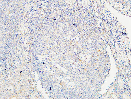
Immunohistochemical analysis of paraffin-embedded Human Amygdala. 1, Antibody was diluted at 1:200(4° overnight). 2, High-pressure and temperature EDTA, pH8.0 was used for antigen retrieval. 3,Secondary antibody was diluted at 1:200(room temperature, 30min).
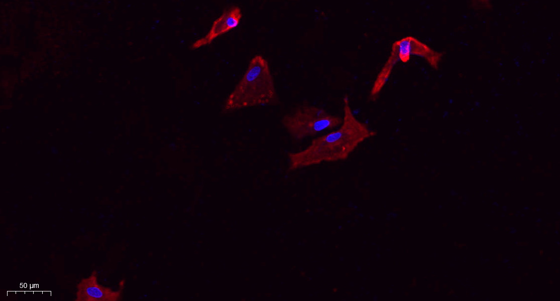
Immunofluorescence analysis of A549. 1,primary Antibody(red) was diluted at 1:200(4°C overnight). 2, Goat Anti Rabbit IgG (H&L) – Alexa Fluor 594 Secondary antibody was diluted at 1:1000(room temperature, 50min).3, Picture B: DAPI(blue) 10min.
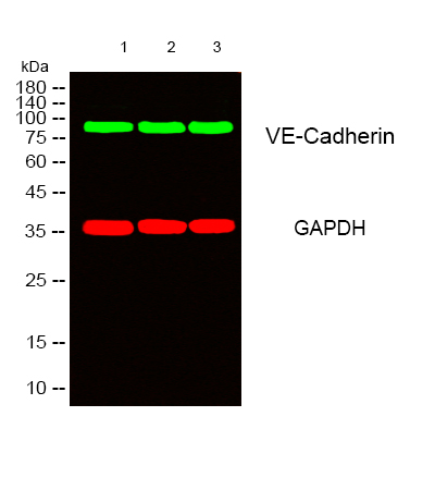
Western blot analysis of lysates from 1) Hela, 2) mouse-lung ,3) mouse-kidney cells, (Green) primary antibody was diluted at 1:1000, 4°over night, secondary antibody(cat:RS23920)was diluted at 1:10000, 37° 1hour. (Red) GAPDH Monoclonal Antibody(2B8) (cat:YM3029) antibody was diluted at 1:5000 as loading control, 4° over night,secondary antibody(cat:RS23710)was diluted at 1:10000, 37° 1hour.
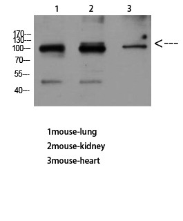
Western Blot analysis of mouse-lung mouse-kidney mouse-heart using VE-Cadherin Polyclonal Antibody diluted at 1:500. Secondary antibody(catalog#:RS0002) was diluted at 1:20000
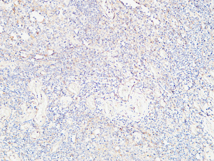
Immunohistochemical analysis of paraffin-embedded Human Amygdala. 1, Antibody was diluted at 1:200(4° overnight). 2, High-pressure and temperature EDTA, pH8.0 was used for antigen retrieval. 3,Secondary antibody was diluted at 1:200(room temperature, 30min).

Western Blot analysis of mouse-liver using VE-Cadherin Polyclonal Antibody diluted at 1:500. Secondary antibody(catalog#:RS0002) was diluted at 1:20000
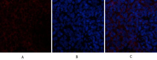
Immunofluorescence analysis of rat-spleen tissue. 1,VE-Cadherin Polyclonal Antibody(red) was diluted at 1:200(4°C,overnight). 2, Cy3 labled Secondary antibody was diluted at 1:300(room temperature, 50min).3, Picture B: DAPI(blue) 10min. Picture A:Target. Picture B: DAPI. Picture C: merge of A+B
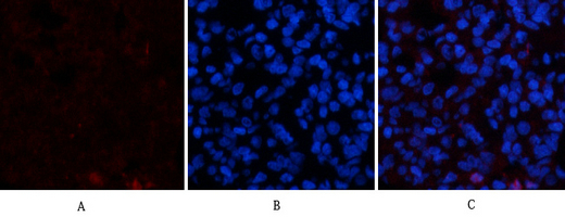
Immunofluorescence analysis of rat-lung tissue. 1,VE-Cadherin Polyclonal Antibody(red) was diluted at 1:200(4°C,overnight). 2, Cy3 labled Secondary antibody was diluted at 1:300(room temperature, 50min).3, Picture B: DAPI(blue) 10min. Picture A:Target. Picture B: DAPI. Picture C: merge of A+B
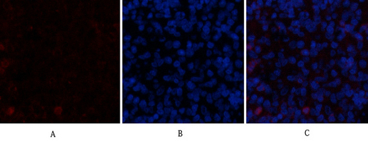
Immunofluorescence analysis of rat-spleen tissue. 1,VE-Cadherin Polyclonal Antibody(red) was diluted at 1:200(4°C,overnight). 2, Cy3 labled Secondary antibody was diluted at 1:300(room temperature, 50min).3, Picture B: DAPI(blue) 10min. Picture A:Target. Picture B: DAPI. Picture C: merge of A+B
For research use only. Not for use in diagnostic procedures.