

Product Name: HMG-1 Polyclonal Antibody
Catalog No.: ALT5502
Reactivity: Human;Mouse;Rat
Applications: IF/ICC;WB;IHC-p;ELISA
Source: Polyclonal, Rabbit,IgG
Formulation: Liquid in PBS containing 50% glycerol, 0.5% BSA and 0.02% sodium azide.
Concentration:1 mg/ml
Dilution: IF: 1:50-200 Western Blot: 1/500 – 1/2000. IHC-p: 1/100-1/300. ELISA: 1/20000. Not yet tested in other applications.
Storage Stability: -20°C/1 year
Gene Name: HMGB1
Protein Name: High mobility group protein B1
Human Gene ID: 3146
Human Swiss Prot No.: P09429
Other Name: HMGB1; HMG1; High mobility group protein B1; High mobility group protein 1; HMG-1
Subcellular Location: Nucleus . Chromosome . Cytoplasm . Secreted . Cell membrane ; Peripheral membrane protein ; Extracellular side . Endosome . Endoplasmic reticulum-Golgi intermediate compartment . In basal state predominantly nuclear. Shuttles between the cytoplasm and the nucleus (PubMed:12231511, PubMed:17114460). Translocates from the nucleus to the cytoplasm upon autophagy stimulation (PubMed:20819940). Release from macrophages in the extracellular milieu requires the activation of NLRC4 or NLRP3 inflammasomes (By similarity). Passively released to the extracellular milieu from necrotic cells by diffusion, involving the fully reduced HGMB1 which subsequently gets oxidized (PubMed:19811284). Also released from apoptotic cells (PubMed:16855214, PubMed:18631454). Active secretion from a variety of immune and non-immune cells such as macrophages, monocytes, neutrophils, dendritic cells and natural killer cells in response to various stimuli such as LPS and cytokines involves a nonconventional secretory process via secretory lysosomes (PubMed:12231511, PubMed:14532127, PubMed:15944249). Secreted by plasma cells in response to LPS (By similarity). Found on the surface of activated platelets (PubMed:11154118). An increased chromatin association is observed when associated with the adenovirus protein pVII (PubMed:27362237). .
Expression: Ubiquitous. Expressed in platelets (PubMed:11154118).
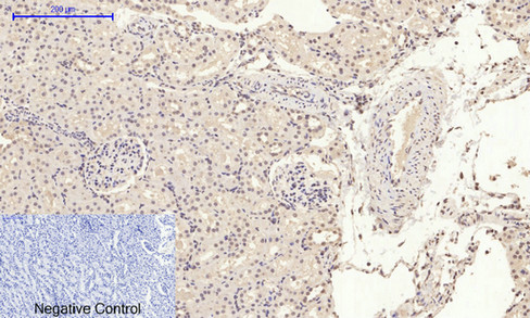
Immunohistochemical analysis of paraffin-embedded Rat-kidney tissue. 1,HMG-1 Polyclonal Antibody was diluted at 1:200(4°C,overnight). 2, Sodium citrate pH 6.0 was used for antibody retrieval(>98°C,20min). 3,Secondary antibody was diluted at 1:200(room tempeRature, 30min). Negative control was used by secondary antibody only.
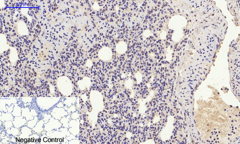
Immunohistochemical analysis of paraffin-embedded Rat-lung tissue. 1,HMG-1 Polyclonal Antibody was diluted at 1:200(4°C,overnight). 2, Sodium citrate pH 6.0 was used for antibody retrieval(>98°C,20min). 3,Secondary antibody was diluted at 1:200(room tempeRature, 30min). Negative control was used by secondary antibody only.
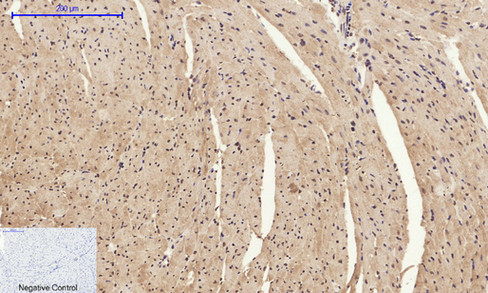
Immunohistochemical analysis of paraffin-embedded Mouse-heart tissue. 1,HMG-1 Polyclonal Antibody was diluted at 1:200(4°C,overnight). 2, Sodium citrate pH 6.0 was used for antibody retrieval(>98°C,20min). 3,Secondary antibody was diluted at 1:200(room tempeRature, 30min). Negative control was used by secondary antibody only.

Immunohistochemical analysis of paraffin-embedded Mouse-lung tissue. 1,HMG-1 Polyclonal Antibody was diluted at 1:200(4°C,overnight). 2, Sodium citrate pH 6.0 was used for antibody retrieval(>98°C,20min). 3,Secondary antibody was diluted at 1:200(room tempeRature, 30min). Negative control was used by secondary antibody only.
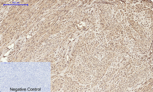
Immunohistochemical analysis of paraffin-embedded Human-uterus tissue. 1,HMG-1 Polyclonal Antibody was diluted at 1:200(4°C,overnight). 2, Sodium citrate pH 6.0 was used for antibody retrieval(>98°C,20min). 3,Secondary antibody was diluted at 1:200(room tempeRature, 30min). Negative control was used by secondary antibody only.
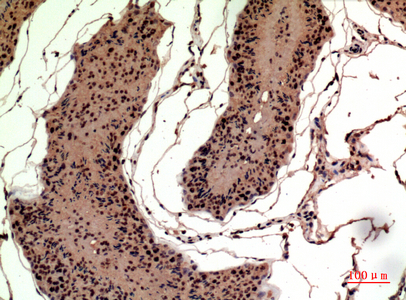
Immunohistochemical analysis of paraffin-embedded rat-testis, antibody was diluted at 1:100
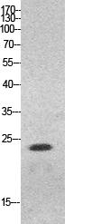
Western Blot analysis of HeLa cells using HMG-1 Polyclonal Antibody. Antibody was diluted at 1:500. Secondary antibody(catalog#:RS0002) was diluted at 1:20000 cells nucleus extracted by Minute TM Cytoplasmic and Nuclear Fractionation kit (SC-003,Inventbiotech,MN,USA).
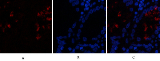
Immunofluorescence analysis of human-stomach tissue. 1,HMG-1 Polyclonal Antibody(red) was diluted at 1:200(4°C,overnight). 2, Cy3 labled Secondary antibody was diluted at 1:300(room temperature, 50min).3, Picture B: DAPI(blue) 10min. Picture A:Target. Picture B: DAPI. Picture C: merge of A+B

For research use only. Not for use in diagnostic procedures.