

Product Name: CD63 Polyclonal Antibody
Catalog No.: ALT5525
Reactivity: Human;Rat;Mouse;
Applications: IF/ICC;WB;IHC-p;ELISA
Source: Polyclonal, Rabbit,IgG
Formulation: Liquid in PBS containing 50% glycerol, 0.5% BSA and 0.02% sodium azide.
Concentration:1 mg/ml
Dilution: IF: 1:50-200 Western Blot: 1/500 – 1/2000. IHC-p: 1:100-1:300. ELISA: 1/20000. Not yet tested in other applications.
Storage Stability: -20°C/1 year
Gene Name: CD63
Protein Name: CD63 antigen
Human Gene ID: 967
Human Swiss Prot No.: P08962
Other Name: CD63; MLA1; TSPAN30; CD63 antigen; Granulophysin; Lysosomal-associated membrane protein 3; LAMP-3; Melanoma-associated antigen ME491; OMA81H; Ocular melanoma-associated antigen; Tetraspanin-30; Tspan-30; CD63
Subcellular Location: Cell membrane ; Multi-pass membrane protein . Lysosome membrane ; Multi-pass membrane protein . Late endosome membrane ; Multi-pass membrane protein . Endosome, multivesicular body . Melanosome . Secreted, extracellular exosome . Cell surface . Also found in Weibel-Palade bodies of endothelial cells (PubMed:10793155). Located in platelet dense granules (PubMed:7682577). Detected in a subset of pre-melanosomes. Detected on intralumenal vesicles (ILVs) within multivesicular bodies (PubMed:21962903). .
Expression: Detected in platelets (at protein level). Dysplastic nevi, radial growth phase primary melanomas, hematopoietic cells, tissue macrophages.
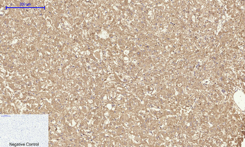
Immunohistochemical analysis of paraffin-embedded Human-kidney-cancer tissue. 1,CD63 Polyclonal Antibody was diluted at 1:200(4°C,overnight). 2, Sodium citrate pH 6.0 was used for antibody retrieval(>98°C,20min). 3,Secondary antibody was diluted at 1:200(room tempeRature, 30min). Negative control was used by secondary antibody only.
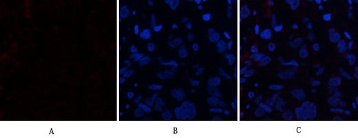
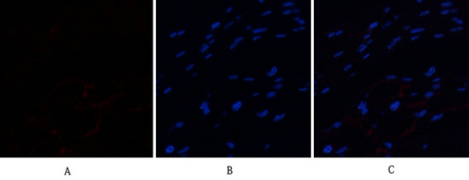
Immunofluorescence analysis of human-breast-cancer tissue. 1,CD63 Polyclonal Antibody(red) was diluted at 1:200(4°C,overnight). 2, Cy3 labled Secondary antibody was diluted at 1:300(room temperature, 50min).3, Picture B: DAPI(blue) 10min. Picture A:Target. Picture B: DAPI. Picture C: merge of A+B
.jpg)
Zhang, Z., Xu, R., Yang, Y. et al. Micro/nano-textured hierarchical titanium topography promotes exosome biogenesis and secretion to improve osseointegration. J Nanobiotechnol 19, 78 (2021).
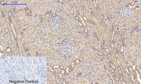
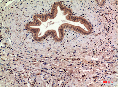
Immunohistochemical analysis of paraffin-embedded Human-kidney tissue. 1,CD63 Polyclonal Antibody was diluted at 1:200(4°C,overnight). 2, Sodium citrate pH 6.0 was used for antibody retrieval(>98°C,20min). 3,Secondary antibody was diluted at 1:200(room tempeRature, 30min). Negative control was used by secondary antibody only.
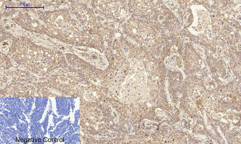
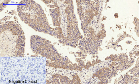
Immunohistochemical analysis of paraffin-embedded Human-lung-cancer tissue. 1,CD63 Polyclonal Antibody was diluted at 1:200(4°C,overnight). 2, Sodium citrate pH 6.0 was used for antibody retrieval(>98°C,20min). 3,Secondary antibody was diluted at 1:200(room tempeRature, 30min). Negative control was used by secondary antibody only.
Immunohistochemical analysis of paraffin-embedded human-liver, antibody was diluted at 1:100
.jpg)
Immunohistochemical analysis of paraffin-embedded human-liver, antibody was diluted at 1:100
Immunohistochemical analysis of paraffin-embedded Human-liver-cancer tissue. 1,CD63 Polyclonal Antibody was diluted at 1:200(4°C,overnight). 2, Sodium citrate pH 6.0 was used for antibody retrieval(>98°C,20min). 3,Secondary antibody was diluted at 1:200(room tempeRature, 30min). Negative control was used by secondary antibody only.
Immunofluorescence analysis of human-stomach-cancer tissue. 1,CD63 Polyclonal Antibody(red) was diluted at 1:200(4°C,overnight). 2, Cy3 labled Secondary antibody was diluted at 1:300(room temperature, 50min).3, Picture B: DAPI(blue) 10min. Picture A:Target. Picture B: DAPI. Picture C: merge of A+B
For research use only. Not for use in diagnostic procedures.