

Product Name: AR Polyclonal Antibody
Catalog No.: ALT5538
Reactivity: Human;Mouse;Rat
Applications: IF/ICC;WB;IHC-p;ELISA
Source: Polyclonal, Rabbit,IgG
Formulation: Liquid in PBS containing 50% glycerol, 0.5% BSA and 0.02% sodium azide.
Concentration:1 mg/ml
Dilution: IF: 1:50-200 WB 1:500-2000, ELISA 1:10000-20000 IHC 1:50-300
Storage Stability: -20°C/1 year
Gene Name: AR
Protein Name: Androgen receptor
Human Gene ID: 367
Human Swiss Prot No.: P10275
Other Name: AR; DHTR; NR3C4; Androgen receptor; Dihydrotestosterone receptor; Nuclear receptor subfamily 3 group C member 4
Subcellular Location: Nucleus . Cytoplasm . Detected at the promoter of target genes (PubMed:25091737). Predominantly cytoplasmic in unligated form but translocates to the nucleus upon ligand-binding. Can also translocate to the nucleus in unligated form in the presence of RACK1. .
Expression: [Isoform 2]: Mainly expressed in heart and skeletal muscle. ; [Isoform 3]: Expressed in basal and stromal cells of the prostate (at protein level).
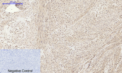
Immunohistochemical analysis of paraffin-embedded Human-uterus tissue. 1,AR Polyclonal Antibody was diluted at 1:200(4°C,overnight). 2, Sodium citrate pH 6.0 was used for antibody retrieval(>98°C,20min). 3,Secondary antibody was diluted at 1:200(room tempeRature, 30min). Negative control was used by secondary antibody only.
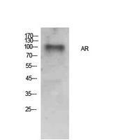
Western Blot analysis of Hela cells using AR Polyclonal Antibody. Secondary antibody(catalog#:RS0002) was diluted at 1:20000
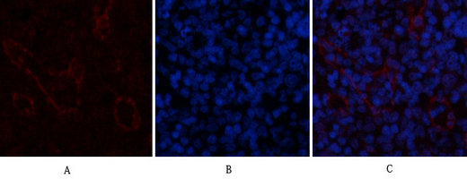
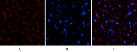
Immunofluorescence analysis of rat-spleen tissue. 1,AR Polyclonal Antibody(red) was diluted at 1:200(4°C,overnight). 2, Cy3 labled Secondary antibody was diluted at 1:300(room temperature, 50min).3, Picture B: DAPI(blue) 10min. Picture A:Target. Picture B: DAPI. Picture C: merge of A+B
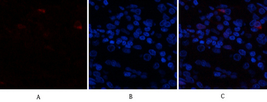
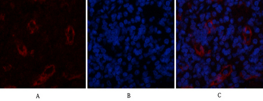
Immunofluorescence analysis of human-stomach tissue. 1,AR Polyclonal Antibody(red) was diluted at 1:200(4°C,overnight). 2, Cy3 labled Secondary antibody was diluted at 1:300(room temperature, 50min).3, Picture B: DAPI(blue) 10min. Picture A:Target. Picture B: DAPI. Picture C: merge of A+B
Immunofluorescence analysis of rat-heart tissue. 1,AR Polyclonal Antibody(red) was diluted at 1:200(4°C,overnight). 2, Cy3 labled Secondary antibody was diluted at 1:300(room temperature, 50min).3, Picture B: DAPI(blue) 10min. Picture A:Target. Picture B: DAPI. Picture C: merge of A+B
Immunofluorescence analysis of rat-spleen tissue. 1,AR Polyclonal Antibody(red) was diluted at 1:200(4°C,overnight). 2, Cy3 labled Secondary antibody was diluted at 1:300(room temperature, 50min).3, Picture B: DAPI(blue) 10min. Picture A:Target. Picture B: DAPI. Picture C: merge of A+B
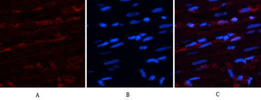
Immunofluorescence analysis of rat-heart tissue. 1,AR Polyclonal Antibody(red) was diluted at 1:200(4°C,overnight). 2, Cy3 labled Secondary antibody was diluted at 1:300(room temperature, 50min).3, Picture B: DAPI(blue) 10min. Picture A:Target. Picture B: DAPI. Picture C: merge of A+B
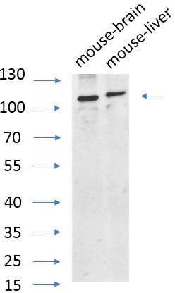
Western Blot analysis of various cells using primary antibody diluted at 1:1000(4°C overnight). Secondary antibody:Goat Anti-rabbit IgG IRDye 800( diluted at 1:5000, 25°C, 1 hour). Cell lysate was extracted by Minute™ Plasma Membrane Protein Isolation and Cell Fractionation Kit(SM-005, Inventbiotech,MN,USA).

For research use only. Not for use in diagnostic procedures.