

Product Name: ABCG2 Polyclonal Antibody
Catalog No.: ALT5558
Reactivity: Human;Rat;Mouse;
Applications: IF/ICC;WB;IHC-p;ELISA
Source: Polyclonal, Rabbit,IgG
Formulation: Liquid in PBS containing 50% glycerol, 0.5% BSA and 0.02% sodium azide.
Concentration:1 mg/ml
Dilution: IF: 1:50-200 Western Blot: 1/500 – 1/2000. IHC-p: 1:100-1:300. ELISA: 1/10000. Not yet tested in other applications.
Storage Stability: -20°C/1 year
Gene Name: ABCG2
Protein Name: ATP-binding cassette sub-family G member 2
Human Gene ID: 9429
Human Swiss Prot No.: Q9UNQ0
Other Name: ABCG2; ABCP; BCRP; BCRP1; MXR; ATP-binding cassette sub-family G member 2; Breast cancer resistance protein; CDw338; Mitoxantrone resistance-associated protein; Placenta-specific ATP-binding cassette transporter; CD338
Subcellular Location: Cell membrane ; Multi-pass membrane protein . Apical cell membrane ; Multi-pass membrane protein . Mitochondrion membrane ; Multi-pass membrane protein . Enriched in membrane lipid rafts. .
Expression: Highly expressed in placenta (PubMed:9850061). Low expression in small intestine, liver and colon (PubMed:9861027). Expressed in brain (at protein level) (PubMed:12958161).
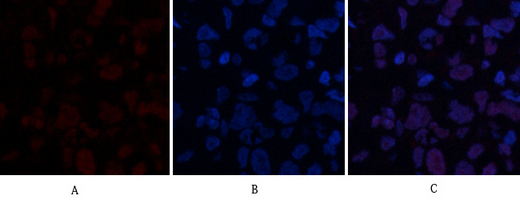
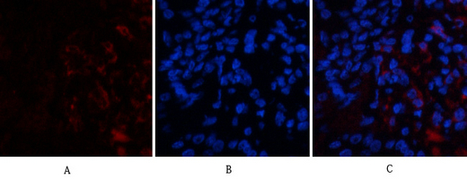
Immunofluorescence analysis of human-breast-cancer tissue. 1,ABCG2 Polyclonal Antibody(red) was diluted at 1:200(4°C,overnight). 2, Cy3 labled Secondary antibody was diluted at 1:300(room temperature, 50min).3, Picture B: DAPI(blue) 10min. Picture A:Target. Picture B: DAPI. Picture C: merge of A+B
Immunofluorescence analysis of human-breast-cancer tissue. 1,ABCG2 Polyclonal Antibody(red) was diluted at 1:200(4°C,overnight). 2, Cy3 labled Secondary antibody was diluted at 1:300(room temperature, 50min).3, Picture B: DAPI(blue) 10min. Picture A:Target. Picture B: DAPI. Picture C: merge of A+B
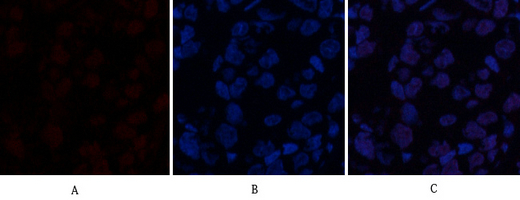
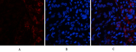
Immunofluorescence analysis of human-lung-cancer tissue. 1,ABCG2 Polyclonal Antibody(red) was diluted at 1:200(4°C,overnight). 2, Cy3 labled Secondary antibody was diluted at 1:300(room temperature, 50min).3, Picture B: DAPI(blue) 10min. Picture A:Target. Picture B: DAPI. Picture C: merge of A+B
Immunofluorescence analysis of human-lung-cancer tissue. 1,ABCG2 Polyclonal Antibody(red) was diluted at 1:200(4°C,overnight). 2, Cy3 labled Secondary antibody was diluted at 1:300(room temperature, 50min).3, Picture B: DAPI(blue) 10min. Picture A:Target. Picture B: DAPI. Picture C: merge of A+B
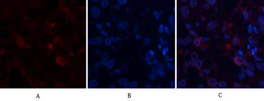
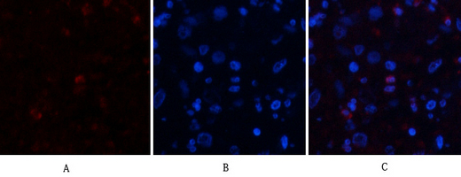
Immunofluorescence analysis of human-kidney tissue. 1,ABCG2 Polyclonal Antibody(red) was diluted at 1:200(4°C,overnight). 2, Cy3 labled Secondary antibody was diluted at 1:300(room temperature, 50min).3, Picture B: DAPI(blue) 10min. Picture A:Target. Picture B: DAPI. Picture C: merge of A+B
Immunofluorescence analysis of human-kidney tissue. 1,ABCG2 Polyclonal Antibody(red) was diluted at 1:200(4°C,overnight). 2, Cy3 labled Secondary antibody was diluted at 1:300(room temperature, 50min).3, Picture B: DAPI(blue) 10min. Picture A:Target. Picture B: DAPI. Picture C: merge of A+B
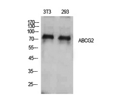
Western Blot analysis of NIH-3T3, 293 cells using ABCG2 Polyclonal Antibody. Antibody was diluted at 1:500. Secondary antibody(catalog#:RS0002) was diluted at 1:20000
.jpg)
Immunohistochemical analysis of paraffin-embedded human-brain, antibody was diluted at 1:100
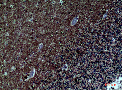
Immunohistochemical analysis of paraffin-embedded human-brain, antibody was diluted at 1:100
For research use only. Not for use in diagnostic procedures.