

Product Name: JIP-1 Polyclonal Antibody
Catalog No.: ALT2434
Reactivity: Human;Mouse;Rat
Applications: WB;IHC-p;IF/ICC;ELISA
Source: Polyclonal, Rabbit,IgG
Formulation: Liquid in PBS containing 50% glycerol, 0.5% BSA and 0.02% sodium azide.
Concentration:1 mg/ml
Dilution: Western Blot: 1/500 – 1/2000. Immunohistochemistry: 1/100 – 1/300. Immunofluorescence: 1/200 – 1/1000. ELISA: 1/5000. Not yet tested in other applications.
Storage Stability: -20°C/1 year
Gene Name: MAPK8IP1
Protein Name: C-Jun-amino-terminal kinase-interacting protein 1
Human Gene ID: 9479
Human Swiss Prot No.: Q9UQF2
Other Name: MAPK8IP1; IB1; JIP1; PRKM8IP; C-Jun-amino-terminal kinase-interacting protein 1; JIP-1; JNK-interacting protein 1; Islet-brain 1; IB-1; JNK MAP kinase scaffold protein 1; Mitogen-activated protein kinase 8-interacting protein 1
Subcellular Location: Cytoplasm . Cytoplasm, perinuclear region . Nucleus . Endoplasmic reticulum membrane. Mitochondrion membrane. Accumulates in cell surface projections. Under certain stress conditions, translocates to the perinuclear region of neurons. In insulin-secreting cells, detected in both the cytoplasm and nucleus (By similarity). .
Expression: Highly expressed in brain. Expressed in neurons, localizing to neurite tips in differentiating cells. Also expressed in the pancreas, testis and prostate. Low levels in heart, ovary and small intestine. Decreased levels in pancreatic beta cells sensitize cells to IL-1-beta-induced apoptosis.
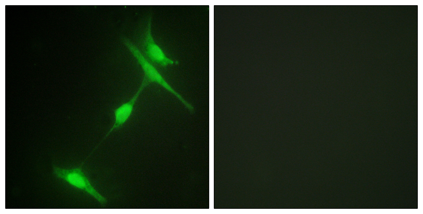
Immunofluorescence analysis of NIH/3T3 cells, using JIP1 Antibody. The picture on the right is blocked with the synthesized peptide.
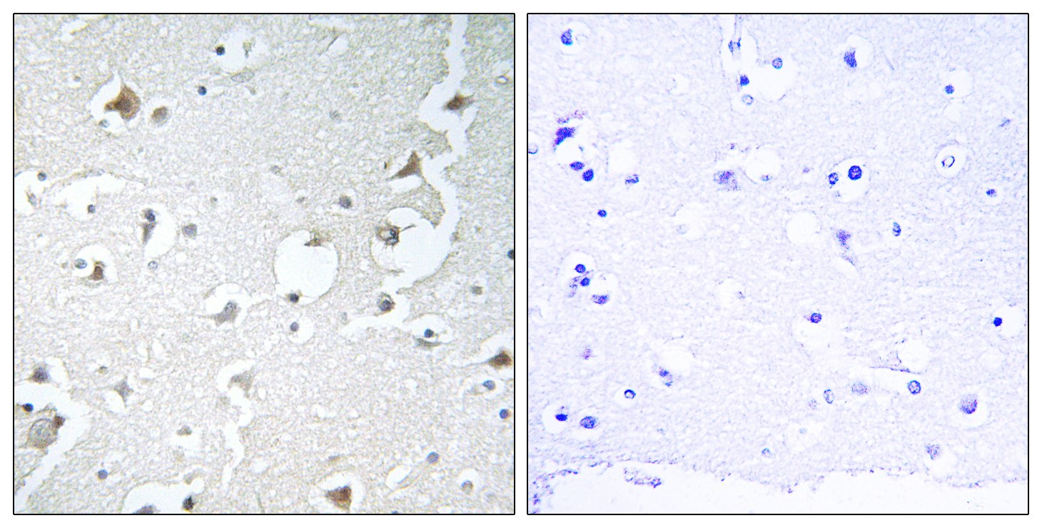
Immunohistochemistry analysis of paraffin-embedded human brain tissue, using JIP1 Antibody. The picture on the right is blocked with the synthesized peptide.
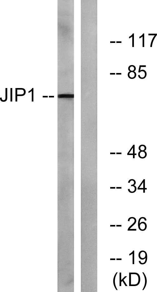
Western blot analysis of lysates from COLO205 cells, treated with Serum 20% 15′, using JIP1 Antibody. The lane on the right is blocked with the synthesized peptide.

For research use only. Not for use in diagnostic procedures.