

Product Name: IRAK-M Polyclonal Antibody
Catalog No.: ALT2393
Reactivity: Human;Rat;Mouse;
Applications: WB;IHC-p;IF/ICC;ELISA
Source: Polyclonal, Rabbit,IgG
Formulation: Liquid in PBS containing 50% glycerol, 0.5% BSA and 0.02% sodium azide.
Concentration:1 mg/ml
Dilution: Western Blot: 1/500 – 1/2000. Immunohistochemistry: 1/100 – 1/300. Immunofluorescence: 1/200 – 1/1000. ELISA: 1/20000. Not yet tested in other applications.
Storage Stability: -20°C/1 year
Gene Name: IRAK3
Protein Name: InteYeukin-1 receptor-associated kinase 3
Human Gene ID: 11213
Human Swiss Prot No.: Q9Y616
Other Name: IRAK3; InteYeukin-1 receptor-associated kinase 3; IRAK-3; IL-1 receptor-associated kinase M; IRAK-M
Subcellular Location: Cytoplasm . Nucleus . In dendritic cells, translocates into the nucleus upon IL33 stimulation. .
Expression: Expressed in eosinophils, dendritic cells and/or monocytes (at protein level) (PubMed:29686383). Expressed predominantly in peripheral blood lymphocytes (PubMed:10383454).
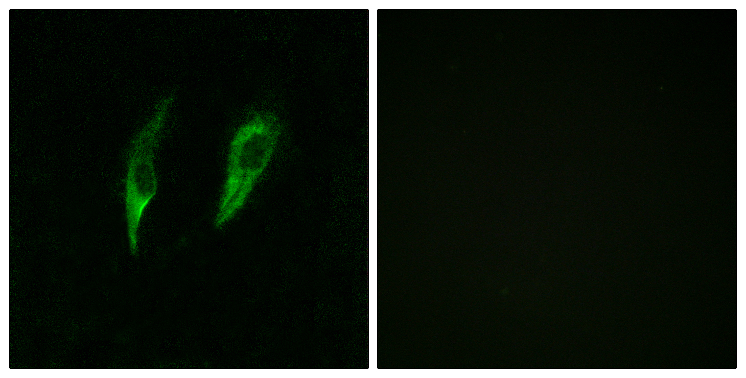
Immunofluorescence analysis of HeLa cells, using IRAK3 Antibody. The picture on the right is blocked with the synthesized peptide.

Immunohistochemistry analysis of paraffin-embedded human brain, using IRAK3 Antibody. The picture on the right is blocked with the synthesized peptide.
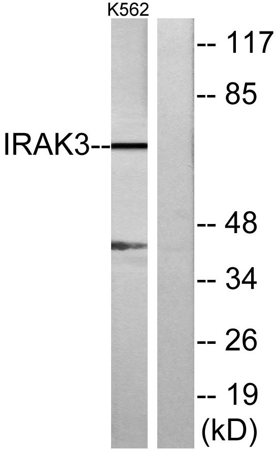
Western blot analysis of lysates from K562 cells, using IRAK3 Antibody. The lane on the right is blocked with the synthesized peptide.
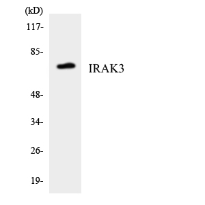
Western blot analysis of the lysates from HUVECcells using IRAK3 antibody.

For research use only. Not for use in diagnostic procedures.