

Product Name: Integrin β1 (phospho Thr789) Polyclonal Antibody
Catalog No.: ALP0879
Reactivity: Human;Mouse;Rat
Applications: WB;IHC-p;IF/ICC;ELISA
Source: Polyclonal, Rabbit,IgG
Formulation: Liquid in PBS containing 50% glycerol, 0.5% BSA and 0.02% sodium azide.
Concentration:1 mg/ml
Dilution: Western Blot: 1/500 – 1/2000. Immunohistochemistry: 1/100 – 1/300. Immunofluorescence: 1/200 – 1/1000. ELISA: 1/5000. Not yet tested in other applications.
Storage Stability: -20°C/1 year
Gene Name: ITGB1
Protein Name: Integrin beta-1
Human Gene ID: 3688
Human Swiss Prot No.: P05556
Other Name: ITGB1; FNRB; MDF2; MSK12; Integrin beta-1; Fibronectin receptor subunit beta; VLA-4 subunit beta; CD antigen CD29
Subcellular Location: Cell membrane ; Single-pass type I membrane protein . Cell projection, invadopodium membrane ; Single-pass type I membrane protein . Cell projection, ruffle membrane ; Single-pass type I membrane protein . Recycling endosome . Melanosome . Cleavage furrow . Cell projection, lamellipodium . Cell projection, ruffle . Cell junction, focal adhesion . Cell surface . Isoform 2 does not localize to focal adhesions. Highly enriched in stage I melanosomes. Located on plasma membrane of neuroblastoma NMB7 cells. In a lung cancer cell line, in prometaphase and metaphase, localizes diffusely at the membrane and in a few intracellular vesicles. In eaYy telophase, detected mainly on the matrix-facing side of the cells. By mid-telophase, concentrated to the ingressing cleavage furrow, mainly to the basal side of the furrow. In late telophase, concentrated to the extending protrusions formed at the opposite ends of the spreading daughter cells, in vesicles at the base of the lamellipodia formed by the separating daughter cells. Colocalizes with ITGB1BP1 and metastatic suppressor protein NME2 at the edge or peripheral ruffles and lamellipodia during the eaYy stages of cell spreading on fibronectin or collagen. Translocates from peripheral focal adhesions sites to fibrillar adhesions in a ITGB1BP1-dependent manner. Enriched preferentially at invadopodia, cell membrane protrusions that correspond to sites of cell invasion, in a collagen-dependent manner. Localized at plasma and ruffle membranes in a collagen-independent manner. .; [Isoform 5]: Cell membrane, sarcolemma . Cell junction . In cardiac muscle, isoform 5 is found in costameres and intercalated disks. .
Expression: [Isoform 1]: Widely expressed, other isoforms are generally coexpressed with a more restricted distribution. ; [Isoform 2]: Expressed in skin, liver, skeletal muscle, cardiac muscle, placenta, umbilical vein endothelial cells, neuroblastoma cells, lymphoma cells, hepatoma cells and astrocytoma cells. ; [Isoform 3]: Together with isoform 4, is expressed in muscle, kidney, liver, placenta, cervical epithelium, umbilical vein endothelial cells, fibroblast cells, embryonal kidney cells, platelets and several blood cell lines. Expressed in non-proliferating and differentiated prostate gland epithelial cells and in platelets, on the surface of erythroleukemia cells and in various hematopoietic cell lines. ; [Isoform 4]: Together with isoform 3, is expressed in muscle, kidney, liver, placenta, cervical epithelium, umbilical vein endothelial cells, fibroblast cells, embryonal kidney cells, platelets and several blood cell lines. Rather than isoform 3, is selectively expressed in peripheral T-cells. ; [Isoform 5]: Expressed specifically in striated muscle (skeletal and cardiac muscle).

Enzyme-Linked Immunosorbent Assay (Phospho-ELISA) for Immunogen Phosphopeptide (Phospho-left) and Non-Phosphopeptide (Phospho-right), using Integrin beta1 (Phospho-Thr789) Antibody

Immunofluorescence analysis of COS7 cells, using Integrin beta1 (Phospho-Thr789) Antibody. The picture on the right is blocked with the phospho peptide.

Immunohistochemistry analysis of paraffin-embedded human breast carcinoma, using Integrin beta1 (Phospho-Thr789) Antibody. The picture on the right is blocked with the phospho peptide.
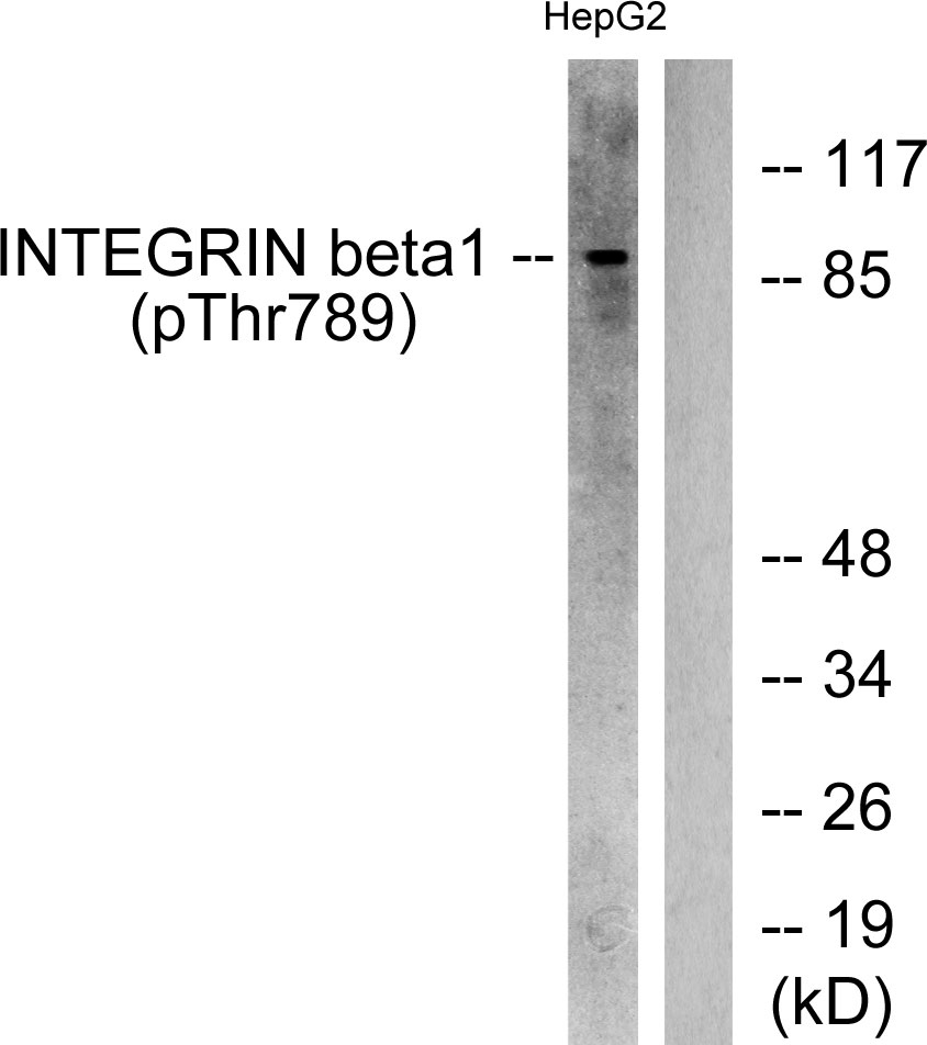
Western blot analysis of lysates from HepG2 cells treated with Ca2+ 40uM 30′, using Integrin beta1 (Phospho-Thr789) Antibody. The lane on the right is blocked with the phospho peptide.

For research use only. Not for use in diagnostic procedures.