

Product Name: Insulin R (phospho Tyr1361) Polyclonal Antibody
Catalog No.: ALP0656
Reactivity: Human;Mouse;Rat
Applications: WB;IHC-p;IF(paraffin section);ELISA
Source: Polyclonal, Rabbit,IgG
Formulation: Liquid in PBS containing 50% glycerol, 0.5% BSA and 0.02% sodium azide.
Concentration:1 mg/ml
Dilution: Western Blot: 1/500 – 1/2000. Immunohistochemistry: 1/100 – 1/300. ELISA: 1/10000. Not yet tested in other applications.
Storage Stability: -20°C/1 year
Gene Name: INSR
Protein Name: Insulin receptor
Human Gene ID: 3643
Human Swiss Prot No.: P06213
Other Name: INSR; Insulin receptor; IR; CD antigen CD220
Subcellular Location: Cell membrane ; Single-pass type I membrane protein . Late endosome . Lysosome . Binding of insulin to INSR induces internalization and lysosomal degradation of the receptor, a means for down-regulating this signaling pathway after stimulation. In the presence of SOY1, internalized INSR molecules are redirected back to the cell surface, thereby preventing their lysosomal catabolism and strengthening insulin signal reception. .
Expression: Isoform Long and isoform Short are predominantly expressed in tissue targets of insulin metabolic effects: liver, adipose tissue and skeletal muscle but are also expressed in the peripheral nerve, kidney, pulmonary alveoli, pancreatic acini, placenta vascular endothelium, fibroblasts, monocytes, granulocytes, erythrocytes and skin. Isoform Short is preferentially expressed in fetal cells such as fetal fibroblasts, muscle, liver and kidney. Found as a hybrid receptor with IGF1R in muscle, heart, kidney, adipose tissue, skeletal muscle, hepatoma, fibroblasts, spleen and placenta (at protein level). Overexpressed in several tumors, including breast, colon, lung, ovary, and thyroid carcinomas.

Enzyme-Linked Immunosorbent Assay (Phospho-ELISA) for Immunogen Phosphopeptide (Phospho-left) and Non-Phosphopeptide (Phospho-right), using IR (Phospho-Tyr1361) Antibody

Immunohistochemistry analysis of paraffin-embedded human breast carcinoma, using IR (Phospho-Tyr1361) Antibody. The picture on the right is blocked with the phospho peptide.
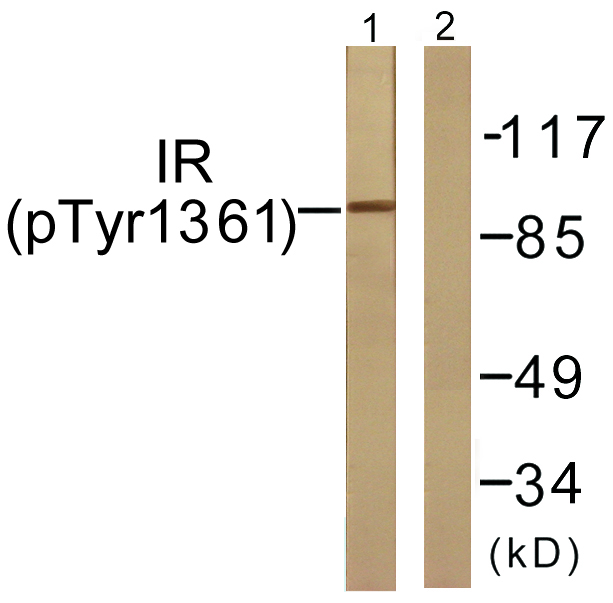
Western blot analysis of lysates from 293 cells treated with Heat shock, using IR (Phospho-Tyr1361) Antibody. The lane on the right is blocked with the phospho peptide.

For research use only. Not for use in diagnostic procedures.