

Product Name: Fes Polyclonal Antibody
Catalog No.: ALT1693
Reactivity: Human;Mouse
Applications: WB;IHC-p;IF(paraffin section);ELISA
Source: Polyclonal, Rabbit,IgG
Formulation: Liquid in PBS containing 50% glycerol, 0.5% BSA and 0.02% sodium azide.
Concentration:1 mg/ml
Dilution: Western Blot: 1/500 – 1/2000. Immunohistochemistry: 1/100 – 1/300. ELISA: 1/20000. Not yet tested in other applications.
Storage Stability: -20°C/1 year
Gene Name: FES
Protein Name: Tyrosine-protein kinase Fes/Fps
Human Gene ID: 2242
Human Swiss Prot No.: P07332
Other Name: FES; FPS; Tyrosine-protein kinase Fes/Fps; Feline sarcoma/Fujinami avian sarcoma oncogene homolog; Proto-oncogene c-Fes; Proto-oncogene c-Fps; p93c-fes
Subcellular Location: Cytoplasm, cytosol. Cytoplasm, cytoskeleton. Cell membrane; Peripheral membrane protein; Cytoplasmic side. Cytoplasmic vesicle. Golgi apparatus. Cell junction, focal adhesion. Distributed throughout the cytosol when the kinase is not activated. Association with microtubules requires activation of the kinase activity. Shuttles between focal adhesions and cell-cell contacts in epithelial cells. Recruited to the lateral cell membrane in polarized epithelial cells by interaction with phosphorylated EZR. Detected at tubular membrane structures in the cytoplasm and at the cell periphery.
Expression: Widely expressed. Detected in adult colon epithelium (at protein level) (PubMed:16455651, PubMed:19051325). Expressed in melanocytes (at protein level) (PubMed:28463229).
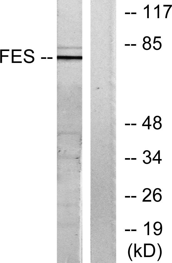
Western blot analysis of lysates from HUVEC cells, treated with serum 20% 30′, using FES Antibody. The lane on the right is blocked with the synthesized peptide.
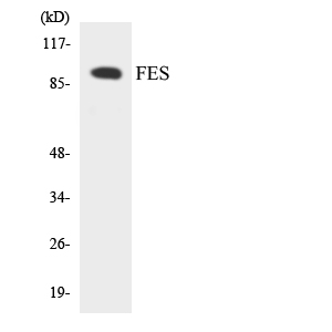
Western blot analysis of the lysates from HUVECcells using FES antibody.

For research use only. Not for use in diagnostic procedures.