

Product Name: EPAS-1 Polyclonal Antibody
Catalog No.: ALT5325
Reactivity: Human;Mouse;Rat
Applications: IF/ICC;WB;IHC-p;ELISA
Source: Polyclonal, Rabbit,IgG
Formulation: Liquid in PBS containing 50% glycerol, 0.5% BSA and 0.02% sodium azide.
Concentration:1 mg/ml
Dilution: IF: 1:50-200 Western Blot: 1/500 – 1/2000. IHC-p: 1/100-1/300. ELISA: 1/20000. Not yet tested in other applications.
Storage Stability: -20°C/1 year
Gene Name: EPAS1 BHLHE73 HIF2A MOP2 PASD2
Protein Name: Endothelial PAS domain-containing protein 1
Human Gene ID: 2034
Human Swiss Prot No.: Q99814
Other Name: EPAS1; BHLHE73; HIF2A; MOP2; PASD2; Endothelial PAS domain-containing protein 1; EPAS-1; Basic-helix-loop-helix-PAS protein MOP2; Class E basic helix-loop-helix protein 73; bHLHe73;HIF-1-alpha-like factor; HLF; Hypoxia-inducible factor 2-alpha; HIF-2-alpha; HIF2-alpha; Member of PAS protein 2; PAS domain-containing protein 2
Subcellular Location: Nucleus . Nucleus speckle . Colocalizes with HIF3A in the nucleus and speckles. .
Expression: Expressed in most tissues, with highest levels in placenta, lung and heart. Selectively expressed in endothelial cells.
.jpg)
Immunohistochemical analysis of paraffin-embedded human-mammary-cancer, antibody was diluted at 1:100
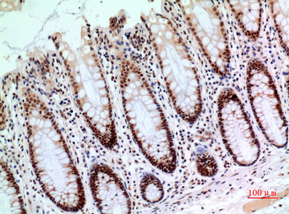
Immunohistochemical analysis of paraffin-embedded human-colon, antibody was diluted at 1:100

Immunofluorescence analysis of human-stomach tissue. 1,EPAS-1 Polyclonal Antibody(red) was diluted at 1:200(4°C,overnight). 2, Cy3 labled Secondary antibody was diluted at 1:300(room temperature, 50min).3, Picture B: DAPI(blue) 10min. Picture A:Target. Picture B: DAPI. Picture C: merge of A+B
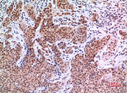
Immunohistochemical analysis of paraffin-embedded human-mammary-cancer, antibody was diluted at 1:100
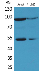
Western Blot analysis of Jurkat, L929 cells using EPAS-1 Polyclonal Antibody. Antibody was diluted at 1:500. Secondary antibody(catalog#:RS0002) was diluted at 1:20000
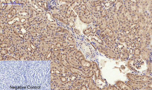
Immunohistochemical analysis of paraffin-embedded Mouse-kidney tissue. 1,EPAS-1 Polyclonal Antibody was diluted at 1:200(4°C,overnight). 2, Sodium citrate pH 6.0 was used for antibody retrieval(>98°C,20min). 3,Secondary antibody was diluted at 1:200(room tempeRature, 30min). Negative control was used by secondary antibody only.
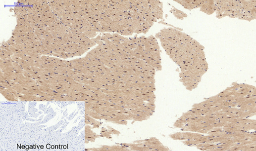
Immunohistochemical analysis of paraffin-embedded Rat-heart tissue. 1,EPAS-1 Polyclonal Antibody was diluted at 1:200(4°C,overnight). 2, Sodium citrate pH 6.0 was used for antibody retrieval(>98°C,20min). 3,Secondary antibody was diluted at 1:200(room tempeRature, 30min). Negative control was used by secondary antibody only.

Immunofluorescence analysis of mouse-spleen tissue. 1,EPAS-1 Polyclonal Antibody(red) was diluted at 1:200(4°C,overnight). 2, Cy3 labled Secondary antibody was diluted at 1:300(room temperature, 50min).3, Picture B: DAPI(blue) 10min. Picture A:Target. Picture B: DAPI. Picture C: merge of A+B
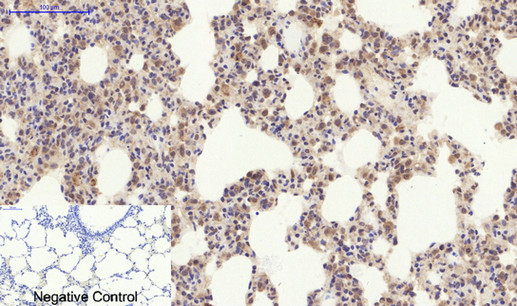
Immunohistochemical analysis of paraffin-embedded Rat-lung tissue. 1,EPAS-1 Polyclonal Antibody was diluted at 1:200(4°C,overnight). 2, Sodium citrate pH 6.0 was used for antibody retrieval(>98°C,20min). 3,Secondary antibody was diluted at 1:200(room tempeRature, 30min). Negative control was used by secondary antibody only.
For research use only. Not for use in diagnostic procedures.