

Product Name: Cdk5 (phospho Tyr15) Polyclonal Antibody
Catalog No.: ALP0380
Reactivity: Human;Mouse;Rat;Monkey
Applications: WB;IHC-p;IF(paraffin section);ELISA
Source: Polyclonal, Rabbit,IgG
Formulation: Liquid in PBS containing 50% glycerol, 0.5% BSA and 0.02% sodium azide.
Concentration:1 mg/ml
Dilution: Western Blot: 1/500 – 1/2000. Immunohistochemistry: 1/100 – 1/300. ELISA: 1/5000. Not yet tested in other applications.
Storage Stability: -20°C/1 year
Gene Name: CDK5
Protein Name: Cyclin-dependent kinase 5
Human Gene ID: 1020
Human Swiss Prot No.: Q00535
Other Name: CDK5; CDKN5; Cyclin-dependent kinase 5; Cell division protein kinase 5; Serine/threonine-protein kinase PSSALRE; Tau protein kinase II catalytic subunit; TPKII catalytic subunit
Subcellular Location: [Isoform 1]: Cytoplasm . Nucleus . Cell membrane ; Peripheral membrane protein. Perikaryon. Cell projection, lamellipodium . Cell projection, growth cone . Cell junction, synapse, postsynaptic density . Cell junction, synapse . In axonal growth cone with extension to the peripheral lamellipodia (By similarity). Under neurotoxic stress and neuronal injury conditions, CDK5R (p35) is cleaved by calpain to generate CDK5R1 (p25) in response to increased intracellular calcium. The elevated level of p25, when in complex with CDK5, leads to its subcellular misallocation as well as its hyperactivation. Colocalizes with CTNND2 in the cell body of neuronal cells, and with CTNNB1 in the cell-cell contacts and plasma membrane of undifferentiated and differentiated neuroblastoma cells. Reversibly attached to the plasma membrane in an inactive form when complexed to dephosphorylated p35 or CDK5R2 (p39), p35 phosphorylation releases this attachment and activates CDK5. .; [Isoform 2]: Nucleus.
Expression: [Isoform 1]: Ubiquitously expressed (PubMed:17009320, PubMed:19693690). Accumulates in cortical neurons (at protein level) (PubMed:17009320). ; [Isoform 2]: Expressed in the testis, skeletal muscle, colon, bone marrow and ovary.
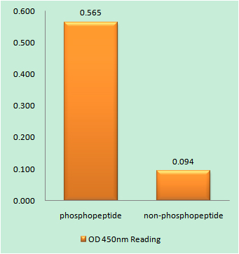
Enzyme-Linked Immunosorbent Assay (Phospho-ELISA) for Immunogen Phosphopeptide (Phospho-left) and Non-Phosphopeptide (Phospho-right), using CDK5 (Phospho-Tyr15) Antibody

Western blot analysis of lysates from COS7 cells treated with EGF 200ng/ml 30′ and 293 cells treated with H2O2 100u, 15mins, using CDK5 (Phospho-Tyr15) Antibody. The lane on the right is blocked with the phospho peptide.

For research use only. Not for use in diagnostic procedures.