Product Name: Catenin-β (phospho Tyr489) Polyclonal Antibody
Catalog No.: ALP0768
Reactivity: Human;Mouse;Rat;Monkey
Applications: WB;IHC-p;IF(paraffin section);ELISA
Source: Polyclonal, Rabbit,IgG
Formulation: Liquid in PBS containing 50% glycerol, 0.5% BSA and 0.02% sodium azide.
Concentration:1 mg/ml
Dilution: Western Blot: 1/500 – 1/2000. Immunohistochemistry: 1/100 – 1/300. ELISA: 1/40000. Not yet tested in other applications.
Storage Stability: -20°C/1 year
Gene Name: CTNNB1
Protein Name: Catenin beta-1
Human Gene ID: 1499
Human Swiss Prot No.: P35222
Other Name: CTNNB1; CTNNB; OK/SW-cl.35; Catenin beta-1; Beta-catenin
Subcellular Location: Cytoplasm . Nucleus . Cytoplasm, cytoskeleton . Cell junction, adherens junction . Cell junction . Cell membrane . Cytoplasm, cytoskeleton, microtubule organizing center, centrosome. Cytoplasm, cytoskeleton, spindle pole. Cell junction, synapse . Cytoplasm, cytoskeleton, cilium basal body . Colocalized with RAPGEF2 and TJP1 at cell-cell contacts (By similarity). Cytoplasmic when it is unstabilized (high level of phosphorylation) or bound to CDH1. Translocates to the nucleus when it is stabilized (low level of phosphorylation). Interaction with GLIS2 and MUC1 promotes nuclear translocation. Interaction with EMD inhibits nuclear localization. The majority of beta-catenin is localized to the cell membrane. In interphase, colocalizes with CROCC between CEP250 puncta at the proximal end of centrioles, and this localization is dependent on CROCC and CEP250. In mitosis, when NEK2 activity increases, it localizes to centrosomes at spindle poles independent of CROCC. Colocalizes with CDK5 in the cell-cell contacts and plasma membrane of undifferentiated and differentiated neuroblastoma cells. Interaction with FAM53B promotes translocation to the nucleus (PubMed:25183871). .
Expression: Expressed in several hair follicle cell types: basal and peripheral matrix cells, and cells of the outer and inner root sheaths. Expressed in colon. Present in cortical neurons (at protein level). Expressed in breast cancer tissues (at protein level) (PubMed:29367600).

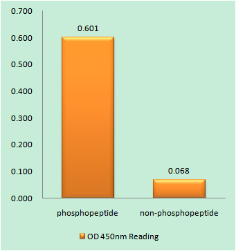
Enzyme-Linked Immunosorbent Assay (Phospho-ELISA) for Immunogen Phosphopeptide (Phospho-left) and Non-Phosphopeptide (Phospho-right), using Catenin-beta (Phospho-Tyr489) Antibody

Immunohistochemistry analysis of paraffin-embedded human brain, using Catenin-beta (Phospho-Tyr489) Antibody. The picture on the right is blocked with the phospho peptide.

Western blot analysis of lysates from COS7 cells treated with UV 15′, using Catenin-beta (Phospho-Tyr489) Antibody. The lane on the right is blocked with the phospho peptide.
For research use only. Not for use in diagnostic procedures.

