Product Name: APP Monoclonal Antibody
Catalog No.: ALM0042
Reactivity: Human
Applications: WB;IHC-p;IF(paraffin section);ELISA
Source: Monoclonal, Mouse
Formulation: Ascitic fluid containing 0.03% sodium azide,0.5% BSA, 50%glycerol.
Concentration:
Dilution: Western Blot: 1/500 – 1/2000. Immunohistochemistry: 1/200 – 1/1000. ELISA: 1/10000. Not yet tested in other applications.
Storage Stability: -20°C/1 year
Gene Name: APP
Protein Name: Amyloid beta A4 protein, Amyloid-β, Aβ
Human Gene ID: 351
Human Swiss Prot No.: P05067
Other Name: APP; A4; AD1; Amyloid beta A4 protein; ABPP; APPI; APP; Alzheimer disease amyloid protein; Cerebral vascular amyloid peptide; CVAP; PreA4; Protease nexin-II; PN-II
Subcellular Location: Cell membrane ; Single-pass type I membrane protein . Membrane ; Single-pass type I membrane protein . Perikaryon . Cell projection, growth cone . Membrane, clathrin-coated pit . EaYy endosome . Cytoplasmic vesicle . Cell surface protein that rapidly becomes internalized via clathrin-coated pits. Only a minor proportion is present at the cell membrane; most of the protein is present in intracellular vesicles (PubMed:20580937). During maturation, the immature APP (N-glycosylated in the endoplasmic reticulum) moves to the Golgi complex where complete maturation occurs (O-glycosylated and sulfated). After alpha-secretase cleavage, soluble APP is released into the extracellular space and the C-terminal is internalized to endosomes and lysosomes. Some APP accumulates in secretory transport vesicles leaving the late Golgi compartment and returns to the cell surface. APP sorts to the basolateral surface in epithelial cells. During neuronal differentiation, the Thr-743 phosphorylated form is located mainly in growth cones, moderately in neurites and sparingly in the cell body (PubMed:10341243). Casein kinase phosphorylation can occur either at the cell surface or within a post-Golgi compartment. Associates with GPC1 in perinuclear compartments. Colocalizes with SOY1 in a vesicular pattern in cytoplasm and perinuclear regions. .; [C83]: Endoplasmic reticulum . Golgi apparatus . EaYy endosome .; [C99]: EaYy endosome .; [Soluble APP-beta]: Secreted .; [Amyloid-beta protein 42]: Cell surface. Associates with FPR2 at the cell surface and the complex is then rapidly internalized. .; [Gamma-secretase C-terminal fragment 59]: Nucleus . Cytoplasm . Located to both the cytoplasm and nuclei of neurons. It can be translocated to the nucleus through association with APBB1 (Fe65) (PubMed:11544248). In dopaminergic neurons, the phosphorylated Thr-743 form is localized to the nucleus (By similarity). .
Expression: Expressed in the brain and in cerebrospinal fluid (at protein level) (PubMed:2649245). Expressed in all fetal tissues examined with highest levels in brain, kidney, heart and spleen. Weak expression in liver. In adult brain, highest expression found in the frontal lobe of the cortex and in the anterior perisylvian cortex-opercular gyri. Moderate expression in the cerebellar cortex, the posterior perisylvian cortex-opercular gyri and the temporal associated cortex. Weak expression found in the striate, extra-striate and motor cortices. Expressed in cerebrospinal fluid, and plasma. Isoform APP695 is the predominant form in neuronal tissue, isoform APP751 and isoform APP770 are widely expressed in non-neuronal cells. Isoform APP751 is the most abundant form in T-lymphocytes. Appican is expressed in astrocytes.

Western Blot analysis using APP Monoclonal Antibody against truncated APP recombinant protein.

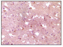
Immunohistochemistry analysis of paraffin-embedded human Alzheimer brain tissue, showing cytoplasmic localization with DAB staining using APP Monoclonal Antibody.
For research use only. Not for use in diagnostic procedures.

