Product Name: AMPKα1 Monoclonal Antibody
Catalog No.: ALM0024
Reactivity: Human;Mouse;Rat;Monkey
Applications: WB;IHC-p;IF/ICC;FCM;ELISA
Source: Monoclonal, Mouse
Formulation: Ascitic fluid containing 0.03% sodium azide,0.5% BSA, 50%glycerol.
Concentration:
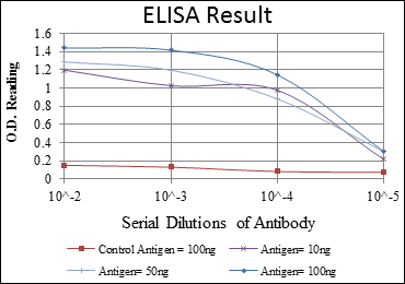
Dilution: Western Blot: 1/500 – 1/2000. Immunohistochemistry: 1/200 – 1/1000. Immunofluorescence: 1/200 – 1/1000. Flow cytometry: 1/200 – 1/400. ELISA: 1/10000. Not yet tested in other applications.
Storage Stability: -20°C/1 year
Gene Name: AAPK1
Protein Name: 5′-AMP-activated protein kinase catalytic subunit alpha-1
Human Gene ID: 5562
Human Swiss Prot No.: Q13131
Other Name: PRKAA1;AMPK1;5′-AMP-activated protein kinase catalytic subunit alpha-1;AMPK subunit alpha-1;Acetyl-CoA carboxylase kinase;ACACA kinase
Subcellular Location: Cytoplasm . Nucleus . In response to stress, recruited by p53/TP53 to specific promoters. .
Expression: Brain,Intestine,Liver,Mammary gland,Platelet,Testis

Western Blot analysis using AMPKα1 Monoclonal Antibody against Jurkat (1), HeLa (2), HepG2 (3), MCF-7 (4), Cos7 (5), NIH/3T3 (6), K562 (7), HEK293 (8), and PC-12 (9) cell lysate.

Immunohistochemistry analysis of paraffin-embedded ovarian cancer (left) and brain tissues (right) with DAB staining using AMPKα1 Monoclonal Antibody.

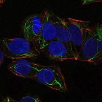
Immunofluorescence analysis of NTERA-2 cells using AMPKα1 Monoclonal Antibody (green). Blue: DRAQ5 fluorescent DNA dye. Red: Actin filaments have been labeled with Alexa Fluor-555 phalloidin.

Flow cytometric analysis of PC-2 cells using AMPKα1 Monoclonal Antibody (green) and negative control (purple).

For research use only. Not for use in diagnostic procedures.

