

Product Name: DRP1 Polyclonal Antibody
Catalog No.: ALT1414
Reactivity: Human;Mouse;Rat
Applications: IF/ICC;WB;IHC-p;ELISA
Source: Polyclonal, Rabbit,IgG
Formulation: Liquid in PBS containing 50% glycerol, 0.5% BSA and 0.02% sodium azide.
Concentration:1 mg/ml
Dilution: IF: 1:50-200 Western Blot: 1/500 – 1/2000. Immunohistochemistry: 1/100 – 1/300. ELISA: 1/10000. Not yet tested in other applications.
Storage Stability: -20°C/1 year
Gene Name: DNM1L
Protein Name: Dynamin-1-like protein
Human Gene ID: 10059
Human Swiss Prot No.: O00429
Other Name: DNM1L; DLP1; DRP1; Dynamin-1-like protein; Dnm1p/Vps1p-like protein; DVLP; Dynamin family member proline-rich carboxyl-terminal domain less; Dymple; Dynamin-like protein; Dynamin-like protein 4; Dynamin-like protein IV; HdynIV; Dynamin-rela
Subcellular Location: Cytoplasm, cytosol. Golgi apparatus. Endomembrane system; Peripheral membrane protein. Mitochondrion outer membrane ; Peripheral membrane protein. Peroxisome. Membrane, clathrin-coated pit . Cytoplasmic vesicle, secretory vesicle, synaptic vesicle membrane . Mainly cytosolic. Recruited by RALA and RALBP1 to mitochondrion during mitosis (PubMed:21822277). Translocated to the mitochondrial membrane through O-GlcNAcylation and interaction with FIS1. Colocalized with MARCHF5 at mitochondrial membrane. Localizes to mitochondria at sites of division. Localizes to mitochondria following necrosis induction. Recruited to the mitochondrial outer membrane by interaction with MIEF1. Mitochondrial recruitment is inhibited by C11orf65/MFI (By similarity). Associated with peroxisomal membranes, partly recruited there by PEX11B. May also be associated with endoplasmic reticulum tubules and cytoplasmic vesicles and found to be perinuclear. In some cell types, localizes to the Golgi complex (By similarity). Binds to phospholipid membranes (By similarity). .
Expression: Ubiquitously expressed with highest levels found in skeletal muscles, heart, kidney and brain. Isoform 1 is brain-specific. Isoform 2 and isoform 3 are predominantly expressed in testis and skeletal muscles respectively. Isoform 4 is weakly expressed in brain, heart and kidney. Isoform 5 is dominantly expressed in liver, heart and kidney. Isoform 6 is expressed in neurons.
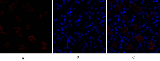
Immunofluorescence analysis of mouse-kidney tissue. 1,DRP1 Polyclonal Antibody(red) was diluted at 1:200(4°C,overnight). 2, Cy3 labled Secondary antibody was diluted at 1:300(room temperature, 50min).3, Picture B: DAPI(blue) 10min. Picture A:Target. Picture B: DAPI. Picture C: merge of A+B
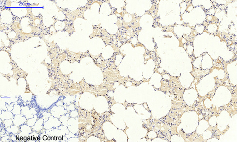
Immunohistochemical analysis of paraffin-embedded Rat-lung tissue. 1,DRP1 Polyclonal Antibody was diluted at 1:200(4°C,overnight). 2, Sodium citrate pH 6.0 was used for antibody retrieval(>98°C,20min). 3,Secondary antibody was diluted at 1:200(room tempeRature, 30min). Negative control was used by secondary antibody only.
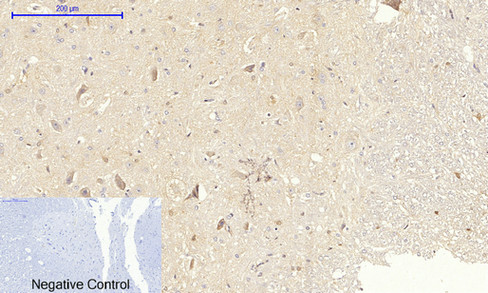
Immunohistochemical analysis of paraffin-embedded Rat-spinal-cord tissue. 1,DRP1 Polyclonal Antibody was diluted at 1:200(4°C,overnight). 2, Sodium citrate pH 6.0 was used for antibody retrieval(>98°C,20min). 3,Secondary antibody was diluted at 1:200(room tempeRature, 30min). Negative control was used by secondary antibody only.

Immunofluorescence analysis of rat-lung tissue. 1,DRP1 Polyclonal Antibody(red) was diluted at 1:200(4°C,overnight). 2, Cy3 labled Secondary antibody was diluted at 1:300(room temperature, 50min).3, Picture B: DAPI(blue) 10min. Picture A:Target. Picture B: DAPI. Picture C: merge of A+B

Immunohistochemical analysis of paraffin-embedded Rat-brain tissue. 1,DRP1 Polyclonal Antibody was diluted at 1:200(4°C,overnight). 2, Sodium citrate pH 6.0 was used for antibody retrieval(>98°C,20min). 3,Secondary antibody was diluted at 1:200(room tempeRature, 30min). Negative control was used by secondary antibody only.
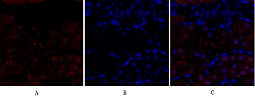
Immunofluorescence analysis of rat-kidney tissue. 1,DRP1 Polyclonal Antibody(red) was diluted at 1:200(4°C,overnight). 2, Cy3 labled Secondary antibody was diluted at 1:300(room temperature, 50min).3, Picture B: DAPI(blue) 10min. Picture A:Target. Picture B: DAPI. Picture C: merge of A+B

Immunohistochemical analysis of paraffin-embedded Mouse-kidney tissue. 1,DRP1 Polyclonal Antibody was diluted at 1:200(4°C,overnight). 2, Sodium citrate pH 6.0 was used for antibody retrieval(>98°C,20min). 3,Secondary antibody was diluted at 1:200(room tempeRature, 30min). Negative control was used by secondary antibody only.
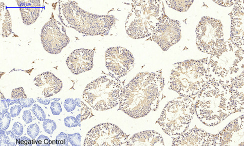
Immunohistochemical analysis of paraffin-embedded Mouse-testis tissue. 1,DRP1 Polyclonal Antibody was diluted at 1:200(4°C,overnight). 2, Sodium citrate pH 6.0 was used for antibody retrieval(>98°C,20min). 3,Secondary antibody was diluted at 1:200(room tempeRature, 30min). Negative control was used by secondary antibody only.

For research use only. Not for use in diagnostic procedures.