

Product Name: p120 (phospho Tyr228) Polyclonal Antibody
Catalog No.: ALP0919
Reactivity: Human;Mouse;Rat
Applications: WB;IHC-p;IF/ICC;ELISA
Source: Polyclonal, Rabbit,IgG
Formulation: Liquid in PBS containing 50% glycerol, 0.5% BSA and 0.02% sodium azide.
Concentration:1 mg/ml
Dilution: Western Blot: 1/500 – 1/2000. Immunohistochemistry: 1/100 – 1/300. Immunofluorescence: 1/200 – 1/1000. ELISA: 1/10000. Not yet tested in other applications.
Storage Stability: -20°C/1 year
Gene Name: CTNND1
Protein Name: Catenin delta-1
Human Gene ID: 1500
Human Swiss Prot No.: O60716
Other Name: CTNND1; KIAA0384; Catenin delta-1; Cadherin-associated Src substrate; CAS; p120 catenin; p120(ctn); p120(cas)
Subcellular Location: Cell junction, adherens junction . Cytoplasm . Nucleus . Cell membrane . Interaction with GLIS2 promotes nuclear translocation (By similarity). Detected at cell-cell contacts (PubMed:15240885, PubMed:17047063). NANOS1 induces its translocation from sites of cell-cell contact to the cytoplasm (PubMed:17047063). CDH1 enhances cell membrane localization (PubMed:15240885). Isoforms 4A and 1AB are excluded from the nucleus (PubMed:11896187). .; [Isoform 1A]: Nucleus .; [Isoform 2A]: Nucleus .; [Isoform 3A]: Nucleus .
Expression: Expressed in vascular endothelium. Melanocytes and melanoma cells primarily express the long isoform 1A, whereas keratinocytes express shorter isoforms, especially 3A. The shortest isoform 4A, is detected in normal keratinocytes and melanocytes, and generally lost from cells derived from squamous cell carcinomas or melanomas. The C-terminal alternatively spliced exon B is present in the p120ctn transcripts in the colon, intestine and prostate, but lost in several tumor tissues derived from these organs.
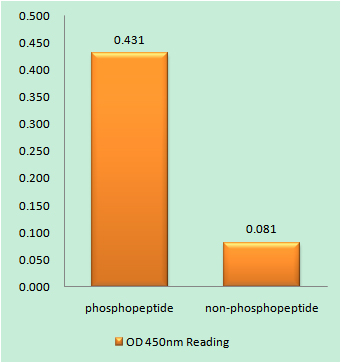
Enzyme-Linked Immunosorbent Assay (Phospho-ELISA) for Immunogen Phosphopeptide (Phospho-left) and Non-Phosphopeptide (Phospho-right), using Catenin-delta1 (Phospho-Tyr228) Antibody

Immunofluorescence analysis of HUVEC cells, using Catenin-delta1 (Phospho-Tyr228) Antibody. The picture on the right is blocked with the phospho peptide.
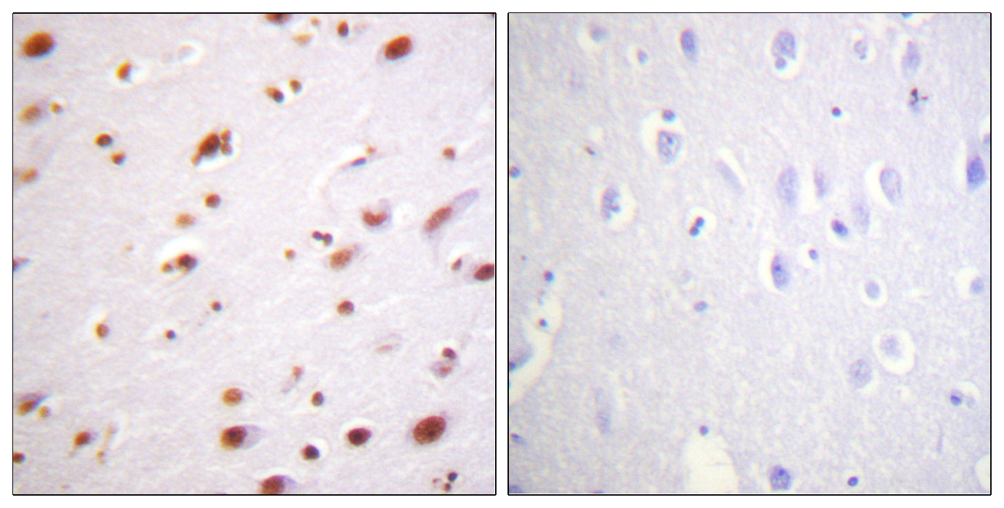
Immunohistochemistry analysis of paraffin-embedded human brain, using Catenin-delta1 (Phospho-Tyr228) Antibody. The picture on the right is blocked with the phospho peptide.
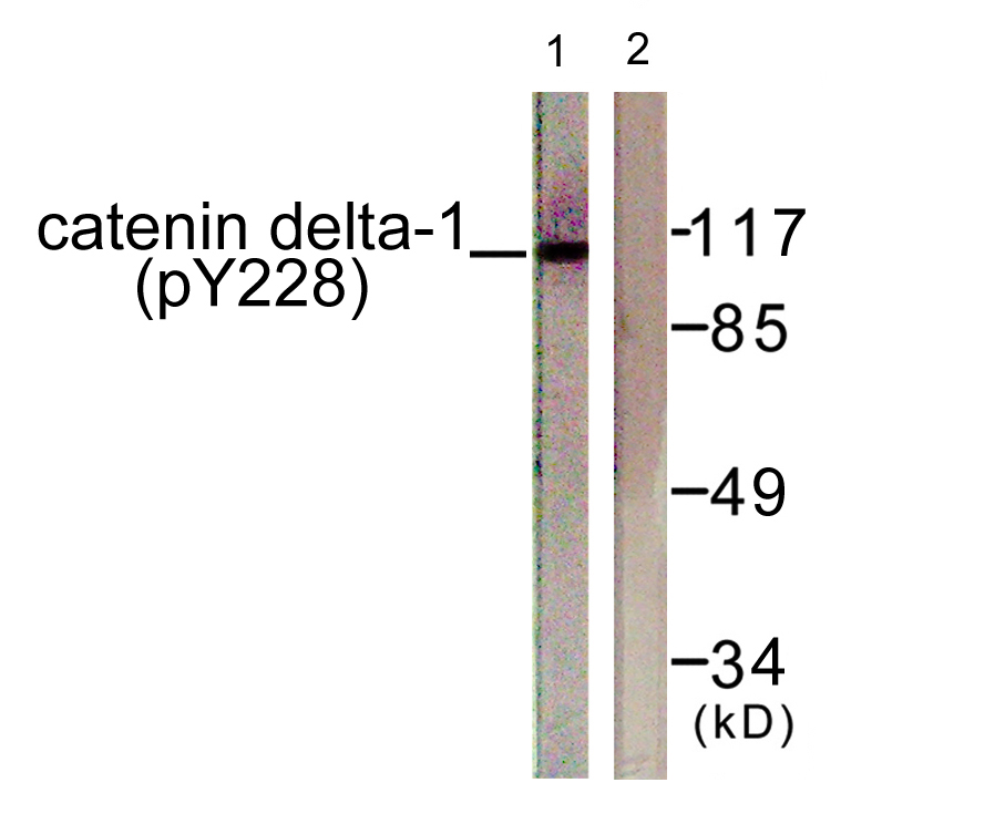
Western blot analysis of lysates from HUVEC cells, using Catenin-delta1 (Phospho-Tyr228) Antibody. The lane on the right is blocked with the phospho peptide.

For research use only. Not for use in diagnostic procedures.