

Product Name: PAKα (phospho Ser199) Polyclonal Antibody
Catalog No.: ALP0757
Reactivity: Human;Mouse;Rat;Monkey
Applications: WB;IHC-p;IF(paraffin section);ELISA
Source: Polyclonal, Rabbit,IgG
Formulation: Liquid in PBS containing 50% glycerol, 0.5% BSA and 0.02% sodium azide.
Concentration:1 mg/ml
Dilution: Western Blot: 1/500 – 1/2000. Immunohistochemistry: 1/100 – 1/300. ELISA: 1/10000. Not yet tested in other applications.
Storage Stability: -20°C/1 year
Gene Name: PAK1
Protein Name: Serine/threonine-protein kinase PAK 1
Human Gene ID: 5058
Human Swiss Prot No.: Q13153
Other Name: PAK1; Serine/threonine-protein kinase PAK 1; Alpha-PAK; p21-activated kinase 1; PAK-1; p65-PAK
Subcellular Location: Cytoplasm . Cell junction, focal adhesion . Cell projection, lamellipodium . Cell membrane . Cell projection, ruffle membrane . Cell projection, invadopodium . Nucleus, nucleoplasm . Chromosome . Cytoplasm, cytoskeleton, microtubule organizing center, centrosome . Colocalizes with RUFY3, F-actin and other core migration components in invadopodia at the cell periphery (PubMed:25766321). Recruited to the cell membrane by interaction with CDC42 and RAC1. Recruited to focal adhesions upon activation. Colocalized with CIB1 within membrane ruffles during cell spreading upon readhesion to fibronectin. Upon DNA damage, translocates to the nucleoplasm when phosphorylated at Thr-212 where is co-recruited with MORC2 on damaged chromatin (PubMed:23260667). Localization to the centrosome does not depend upon the presence of gamma-tubulin (PubMed:27012601). Localization of the active, but not inactive, protein to the adhesions and edge of lamellipodia is mediated by interaction with GIT1 (PubMed:11896197). .
Expression: Overexpressed in gastric cancer cells and tissues (at protein level) (PubMed:25766321).
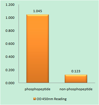
Enzyme-Linked Immunosorbent Assay (Phospho-ELISA) for Immunogen Phosphopeptide (Phospho-left) and Non-Phosphopeptide (Phospho-right), using PAK1 (Phospho-Ser199) Antibody
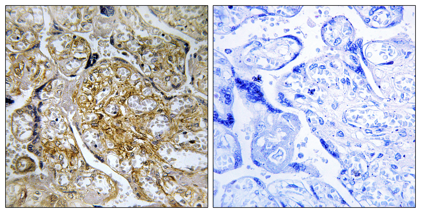
Immunohistochemistry analysis of paraffin-embedded human placenta, using PAK1 (Phospho-Ser199) Antibody. The picture on the right is blocked with the phospho peptide.

Western blot analysis of lysates from LOVO cells treated with starved 24h, using PAK1 (Phospho-Ser199) Antibody. The lane on the right is blocked with the phospho peptide.

For research use only. Not for use in diagnostic procedures.